|
||||||
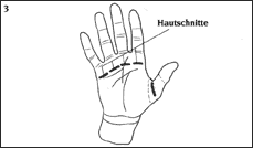
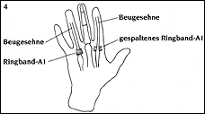
| Definition of Trigger Finger: (also known as Schnappfinger, stenosing tendovaginitis, Digitus saltans, Trigger finger, Snapping finger, Doigt à ressort, Dito a scatto) This is one of the most common hand conditions, which involves a painful catching phenomenon in one or more fingers. The majority of those affected are women over the age of fifty. Meist ist der Daumen betroffen, es folgen der Mittelfinger und dann die übrigen Langfinger. Usually it is the thumb that is affected, but it can also affect the middle finger and the other fingers. The word-for-word translation of "stenosing tendovaginitis" is "constricting inflammation of the tendon sheath ", which was first described by Notta (1850). Tendon sheaths surround extensor and flexor tendons of the hands, wrists, feet and ankles. Tendons are strong, fibrous bands that connect the muscles of the forearm with the bones of the wrist, palm and fingers. This "muscle-tendon system" enables the stretching and bending of the wrist and fingers. Tendons slide through a protective tunnel called the tendon sheath, which consists of a strong outer layer and a delicate inner layer (synovium). The synovium prevents the build up of friction during the gliding of the flexor and extensor tendons. So-called pulleys and cruciate ligaments are located along the finger with the role of fixing the flexor tendons to the bone in order to prevent them from lifting up during active flexing of the distal portion of the palm and fingers. (Illustration1) 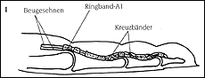 Trigger finger is typically caused by an inflammation-induced narrowing of the tendon sheath, as a rule in the area of the first annular pulley proximal to the finger basal joint. The swelling of a tendon can also cause this catching phenomenon to occur. The synovium of the tendon sheath can become very swollen when inflamed, so that the tendon can only glide with great difficulty or not at all. Friction is then evident and in advanced stages a catching sensation is experienced. During firm bending of the thumb or finger, the swollen portion of the tendon sheath is pulled towards the body and the passing through the narrow opening is often accompanied by a painful popping. The swollen portion of the tendon remains in front of the point of constriction (Illustration 2), 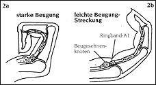 The finger is stuck in the bending position and can only be straightened while experiencing more pain and more popping. Often the patient uses the healthy hand in order to passively straighten the affected finger. Causes of Trigger Finger The first annular pulley is inherently narrow, creating a tendency for this condition to develop under unfavorable conditions, such as repetitive overuse during work or hobbies. Activities that require a firm grip or the extensive use of tools that press against the first annular pulley and other parts of the tendon sheath can irritate the tendon and cause the swelling of the tendon and/or the tendon sheath. The symptoms of trigger finger can also occur as a result of polyarthritis (rheumatic arthritis), gout or other metabolic disorders such as diabetes mellitus. In children the cause of the condition (Pollex flexus congenitus = born with a bent thumb) is primarily due to an alteration of the tendons which are too thick and create a subsequent reaction in the tendon sheath. Signs and Symptoms of Trigger Finger Before the typical popping arises, pain is experienced during the bending of the affected finger in the area of the constriction, that is in the first annular pulley in the distal portion of the palm. Pressure applied to the examined finger results in intensified pain. In early stages of the condition, the gliding and rubbing of the flexor tendons can be felt. Often, a small knot in the area of the tendon in front of the first annular pulley can also be noticed. In advanced cases, the affected finger remains blocked in its bent position, or sometimes, though rarely, in a straightened position. Patients often experience the snapping to be localized in the area of the thumb joint and sometimes this joint is fixed in either a flexed or extended position. The cause lies in the area of the first annular pulley near the basal joint. Diagnosis of Trigger Finger As a rule, the typical case history (anamnesis) and a clinical examination with the determination of the described symptoms leads to a reliable diagnosis. In order to rule out any bone-related causes of the ailments or calcification of soft tissues, an x-ray examination should be performed. In cases of children, the parents or the physician notice that the thumb is fixed in either a bent or straightened position. The child cries when attempts are made to passively straighten the thumb and it remains in a bent position. Treatment of Trigger Finger
There are various possibilities for ensuring that the patient is free of pain during the surgery. These possibilities will be explained to you by the anesthesiologist. Technical Aspects of the Surgery Surgery for trigger finger is usually out-patient, that is, the patient can go home once the operation is completed. 1. General surgical preparation:
Pain as a result of the incision is typically minimal and most patients do not require pain medication. The snapping symptoms and the resulting pain disappear after the surgery although occasionally a rubbing sensation in the tendons is felt which goes away after a few weeks. Scar discomfort largely disappears after the first 6-8 weeks. After 3-6 months patients no longer complain of pain. However, the scars final state is not reached until approximately 12 months after surgery. © Dr. Klaus Lowka |
||||
| Home | About Dr. Lowka | Advanced Education/Publications | The Hand Hand Surgery Diagnosis and Operations | Clinical Pictures in Hand Surgery | Areas of Emphasis in Hand Surgery Post-operative Treatment | Center for Diagnosis and Out-patient Surgery Contact Us | Legal Information |
||||