|
Standard sequence of events in
hand surgery diagnosis
- Medical history:
Just as in all other medical disciplines, an exact medical history is necessary in the evaluation of disease or
injury of the hand. Many potential diagnoses can be posed or narrowed down through a medical history enquiry alone
(for example, the onset and type of ailments, the time of the accident and course of events, previously received
treatments, age of patient, occupation).
- Clinical examination:
With a standardized and exact clinical examination it is possible to provide a hand surgical diagnosis prior to the
use of additional diagnostic methods.
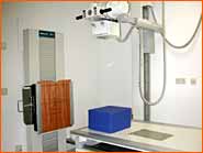
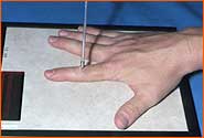
- X-ray examination:
This is part of every hand surgery diagnosis! In order to properly evaluate the x-ray results of one side, the
x-rays of the healthy side should also be taken for comparison!
Every patient with an extremity injury (arm or leg)
must be x-rayed because only in this way is it possible rule out the existence of a fracture! Neglecting to do an
x-ray examination with the comment "Nothing will be broken" is, from my viewpoint and my experience, not permissible.
Overlooked fractures often result in situations where treatment or work disability compensation is more costly than
the correct initial diagnosis with x-ray examination and the subsequent correct therapy.
- Neurological examination:
In cases where nerve lesions are suspected after injury or a syndrome involving etrapment neuropathy is expected,
a neurological exam is required in order to make an exact diagnosis and to plan the appropriate therapy. After surgeries
affecting nerve tissue, the success of the surgery is controlled and monitored through additional neurological exams.
- Computer Tomography (CT):
CT is a special x-ray technique that results in cross-sections of body parts that, through computer reconstructions, create
three-dimensional images of bones. The CT examination is especially useful in the assessment of bone structures. Fractures
can be seen that are missed with conventional x-rays. In addition, the condition of articular surfaces or rotational defects
of bones can be evaluated for surgical planning.
"CT examination of wrist with fractures (break) of the capitate bone (arrow 1) and treiquetral bone (arrow 2)."
- Magnetic resonance imaging (MRT, MRI, NMR):
MRI is a modern imaging technique that does not require x-ray radiation. The utilization of strong magnetic fields aligns
oscillating atoms, thereby absorbing energy.
Elimination of the magnetic field causes this energy to be released in the form
of electromagnetic waves, which are then recorded. Magnetic resonance imaging has developed into a valuable complement to
computer tomography, especially in the diagnosis of changes in soft tissues. With respect to hand surgery, it is especially
useful in the diagnosis of soft tissue tumors, chronic polyarthritis and the assessment of the vitality of bones.
"MRI examination of the wrist and carpus with evidence of a necrosis (tissue death) of the lunate bone (arrow)."
- Radionucleotide imaging:
(also scintiscanning which translates to 'recording of fibrillation')Radio nucleotides (with the shortest possible half-lives) are recorded after intravenous
injection into various tissues. This technique is particularly well-known for its use in the diagnosis of thyroid disease. With
respect to hand surgery, this technique is used to evaluate infected bone and joint changes (arthritis, rheumatoid arthritis) and
the diagnosis of Morbus Sudeck or reflex sympathetic dystrophy (SRD, CRPS I).
Abnormal (pathological) changes of the bone
(skeleton) in the form of a bone growth (osteoplasy) or bone mass loss (osteolysis) can be seen with the aid of radionucleotide
imaging as entrance into the described areas where bone mass was lost is considered a positive test result. This enables a very
early detection of changes in bone mass, in contrast to x-rays which only show bone loss after a 30-50% decrease in calcification.
Radionucleotide imaging = very sensitive, less specific
X-rays = less sensitive, more specific
A very good depiction of the joint is also possible. The injection of a radioactive substance also shows the blood circulation of the region
around a joint or bone (such as in the early phase and perfusion phase) as well as, if required, all metabolic steps (blood pooling phase)
without additional radiation exposure. The radiation exposure is very low now that technetium is generally used as the contrast agent.
1)  2) 2) 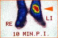
1) "Radionucleotide imaging of both hands with clear accumulation of radionucleotides with reflex sympathetic dystrophy (SRD, CRPS I, Morbus Sudeck).
The arrows refer to the finger joints and the wrist joint."
2) "Radionucleotide imaging of both feet with clear accumulation in area of the upper ankle after a fracture (arrow)."
 back to top back to top
|
|
 |